척주 측만곡증(척주 측만증), Scoliosis
척주 측만곡증(척주 측만증)의 원인
- 척주에 생리적으로 경추 만곡, 흉추 만곡, 요추 만곡 그리고 골반 만곡이 약간 있는 것이 정상적이다.
- 척주의 일부분이 비정상적으로 좌측이나 우측으로 만곡된 상태를 척주 측만이라 한다.
- 척주 측만으로 생기는 증상 징후를 척주 측만증 또는 척주 측만곡증이라 한다.
- 학령기 아이들에게 척주 측만증이 생기는 빈도는 거의 10%이다.
- 그 중 약 60~80%는 특발성 척주 측만증이다.
- 척주 측만증에는 선천성으로 생기는 선천성 척주 측만증과 후천성으로 생기는 후천성 척주 측만증이 있다.
- 원인을 확실히 알 수 없이 생기는 원발성 척주 측만증(특발성 척주 측만증),
- 근육신경 이상 등으로 생기는 근육신경 이상 척주 측만증도 있다.
- 평소에 자세를 바르게 취하지 않아 생기는 척주 측만증,
- 척주염이나 척주외상이나 척주 종양 등으로 척주가 비정상으로 좌측이나 우측으로 조금 만곡된 척주 측만증도 있다.
- 원발성 척주 측만증은 생후 어느 때든지 생길 수 있지만 사춘기가 시작되기 바로 전 부터 더 잘 생길 수 있고
- 여아의 경우 11세경, 남아의 경우 12~14 경에 현저하게 나타나는 것이 보통이다.
- 즉 사춘기가 시작하기 전 약간 만곡되어 있던 척주가 사춘기가 오기 시작하면서 척주 만곡이 불과 1~2년 동안 현저하게 심하게 만곡되어 척주 측만의 중증도가 뚜렷하게 나타날 수 있고 증상 징후도 생길 수 있다.
- 뇌성마비, 소아마비, 척수수막류, 프리드라히 운동실조, 척수외상, 신경 섬유증, 마르팡증, 엘러스단로스증, 척수종양, 척주골종, 구루병, 불완전 골형성증, 비타민 A과다증, 갑상선 기능저하증, 연소성 류마토이드 관절염, 뮤코다당체 침착증 (점다당질증), 척주염, 척주결핵 등으로 척주 측만증이 생길 수 있다.
척주 측만곡증(척주 측만증)의 증상 징후
- 척주 측만증의 원인과 정도에 따라 증상 징후가 다르다.
- 이론적으로 척주의 어느 부위에도 생길 수 있다.
- 흉부 척주에만 생길 수도 있고 흉부 척주의 일부와 허리 척주(요척주)의 일부에만 생길 수도 있고,
- 어떤 때는 허리 척주에만 생길 수 있다.
- 척주 측만이 생긴 척주 부위가 좌측으로 또는 우측으로 경미하게 만곡될 수 있고, 또는 심하게 만곡될 수 있다.
- 척주 측만의 정도가 심하지 않을 때는 척주가 조금 측만 되었을 뿐 아무 증상 징후가 나타나지 않는다.
- 척주 측만이 심하게 생겼을 때는 사진 129, 130과 같이 외관상으로 척주가 측만된 것을 쉽게 알 수 있다.
- 더 심하면 옷을 입고 있는 상태에서 척주가 측만 된 것이 겉으로도 나타나고 평소에 운동을 자유자재로 할 수 없다.
- 심한 척주 측만을 적절히 치료해 주지 않으면 수명이 짧아진다.
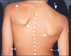
사진 129. 척주 측만.
하얀 점선으로 표시된 선과 같이 척주가 측만되어 있다. 팔과 허리 사이의 간격에 차이가 난다. 양쪽 어깨 높이에 차이가 난다.
Copyright ⓒ 2011 John Sangwon Lee, M.D., FAAP
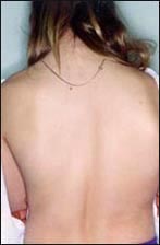
사진 130. 척주 측만이 되어 있다. 사춘기 여아의 후면 등 사진
Copyright ⓒ 2011 John Sangwon Lee, M.D., FAAP
척주 측만곡증(척주 측만증)의 진단
- 척주 측만곡의 중증도에 따라 경도, 중등도, 중증 척주 측만으로 구분한다.
- 병력, 증상 징후와 진찰소견 등을 종합해 척주가 측만되어 있다고 의심되면 척주 측만곡 측정 기구(사진 132. 참조)로 척주 측만의 중증도를 재보고 필요에 따라 척주 X선 사진 검사로 확진한다.
- 경도 척주 측만이 있는 아이들이 옷을 입고 있을 때는 이 병이 있는지 쉽게 알 수 없지만, 옷을 벗고 반듯이 서 있을 때 머리끝에서 엉덩이까지의 척주 전체를 뒤에서 살펴보면 척주의 일부가 우측으로나 좌측으로 휘어지고 한쪽 어깨 높이가 다른 쪽 어깨 높이보다 더 낮다.
- 한쪽 앞가슴이 다른 쪽 앞가슴보다 앞으로 더 불쑥 나와 있다.
- 반듯이 서서 두 팔을 양쪽 옆구리로 반듯이 내려붙일 때 한쪽 옆구리와 그쪽 팔 사이의 간격이 다른 쪽의 사이의 간격보다 더 많이 벌어져서 양쪽 옆구리와 팔 사이의 간격 차이가 다르다.
- 척주 측만곡 측정 기구로 만곡의 정도를 쟀을 때 척주가 6~7도 이상 만곡되어 있으면 척주 만곡이 확실히 있는 것으로 간주한다.
- 마지막으로 척주 X선 사진 검사로 척주 측만의 정도를 더 확실히 알아보고, 또 수술로 치료를 해야 하는지 알아본다.
그림 134.쉬우어만 병으로 생긴 척주 후만증
Copyright ⓒ 2011 John Sangwon Lee, M.D., FAAP
그림 131. 아담스 테스트
양쪽 어깨 높이에 차이가 나고, 뒤에서 등을 볼 때 척주가 왼쪽으로 또는 오른쪽으로 만곡되어 있고, 양쪽 견갑골의 높이에 차이가 나고 양 옆구리 아래로 쭉 내린 각 팔과 옆구리의 사이의 공간의 간격 크기에 차이가 난다.
Copyright ⓒ 2011 John Sangwon Lee, M.D., FAAP
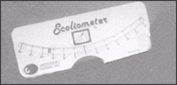
사진 132. 척추 측만곡의 정도를 재는 기구
Copyright ⓒ 2011 John Sangwon Lee, M.D., FAAP
척주 측만곡증(척주 측만증)의 치료
- 척주 측만곡의 정도에 따라 치료가 다르다.
- 경도 척주 측만이 있을 때는 자신도 가족들도 척주 측만곡이 있는지 잘 알지 못할 때가 많다.
- 그렇지만 경도 척주 측만이 불과 몇 달 동안 중증 척주 측만곡으로 더 나빠질 수 있다.
- 정기 건강검진을 받을 때마다 척주 측만이 있는지 통상적으로 알아보아야 한다.
- 경도 척주 측만을 조기에 진단해서 더 이상 악화되지 않도록 조기에 적절히 치료해야 한다.
- 경도 척주 측만은 적절한 운동과 물리치료 등으로 치료한다.
- 경증 척주 측만을 처음 진단 받았을 때부터 적어도 만 18세가 될 때까지 더 악화되는지 알아보기 위해 주기적으로 진찰 받고 필요에 따라 적절한 치료도 받아야 한다.
- 경도 척주 측만이 짧은 기간 동안 더 악화되어 중등도나 중증 척주 측만으로 진행될 수 있다.
- 그때그때 척주 측만의 진행 정도에 따라 운동이나 물리치료나 찰스톤(Charleston) 브레이스 등으로 적기에 적절히 치료받아야 한다.
- 이렇게 치료해서 될 수 있는 한 수술치료를 피해야 한다.
- 중증 척주 측만은 밀와키 브레이스 또는 척수 수술 등으로 치료한다.
- 브레이스 치료를 해줄 때는 목에서부터 엉덩이까지 걸치는 브레이스로 2~4년간 밤낮 치료를 하거나 찰스톤(Charleston) 브레이스로 낮에만 치료할 수 있다.
- 선천성 척주 측만은 3~4세에 수술로 치료할 수 있고
- 근육신경성 척주 측만증은 브레이스 치료로 잘 치료되지 않고 수술로 치료하는 것이 일반적이다.
- 오랫동안 이런 치료를 받을 때 육체적으로 정신적으로 큰 부담이 간다.
- 그러므로 척주 측만은 조기에 진단하여 가능한 한 운동치료나 물리치료 등 비 침입성 치료로 더 쉽고 덜 고생하게 치료해야 한다.
- 즉 브레이스 치료나 척수 수술 치료를 꼭 해야 할 정도까지 척주 측만이 진행되지 않도록 최대한 노력을 해야 한다.
- 중기나 후기 사춘기 아이들에 생기는 특발성 척주 측만의 예후는 일반적으로 좋다.
Scoliosis
Causes of scoliosis
• It is normal for the spine to have physiological cervical, thoracic, lumbar, and pelvic curvature.
• A condition in which a part of the spine is abnormally curved to the left or right is called scoliosis.
• Symptomatic signs of scoliosis are called scoliosis or scoliosis.
• The incidence of scoliosis in school-age children is nearly 10%.
• About 60-80% of them are idiopathic scoliosis.
• There are two types of scoliosis: congenital scoliosis and acquired scoliosis. • primary scoliosis of unknown cause (idiopathic scoliosis);
• There is also scoliosis, which is caused by muscular nerve abnormalities such as muscular nerve abnormalities.
• Scoliosis caused by improper posture,
• There is also scoliosis, in which the spine is slightly curved to the left or right due to spondylitis, spinal trauma, or a tumor in the spine.
• Primary scoliosis can develop at any time after birth, but is more likely just before the onset of puberty and
• It is usually noticeable around the age of 11 in girls and around 12-14 in boys.
• In other words, the spinal column, which was slightly curved before the onset of puberty, becomes significantly more curved for only 1 to 2 years as puberty begins.
• Cerebral palsy, polio, meninges, Friedrich’s ataxia, spinal cord trauma, neurofibrosis, marfanosis, elasdanlososis, spinal tumor, osteosarcoma, rickets, osteogenesis imperfecta, hypervitaminosis A, hypothyroidism, Scoliosis can be caused by juvenile rheumatoid arthritis, mucopolysaccharide deposition (point polysaccharidosis), spondylitis, and spinal tuberculosis.
Signs, symptoms of scoliosis
• Symptoms and signs vary depending on the cause and severity of scoliosis.
• Theoretically, it can occur anywhere in the spine.
• It may occur only in the thoracic vertebrae or only part of the thoracic vertebrae and parts of the lumbar vertebrae (radiovertebrae);
• Sometimes it can only occur in the lumbar spine.
• The area of the spinal column with scoliosis may be slightly curved to the left or right, or it may be severely curved.
• When the degree of scoliosis is not severe, there is only a slight scoliosis and there are no symptoms.
• When the scoliosis is severe, it is easy to see that the spinal column is scoliosis, as shown in pictures 129 and 130.
• In more severe cases, a scoliosis of the spine while wearing clothes is apparent, and it is not possible to exercise freely.
• If severe scoliosis is not properly treated, life expectancy is shortened.
Photo 129. Scoliosis of the spine. As shown by the white dotted line, the spinal column is scoliosis. There is a difference in the distance between the arm and the waist. There is a difference in the height of both shoulders. Copyright ⓒ 2011 John Sangwon Lee, M.D., FAAP
Picture 130. Only the vertebral column is shown. Back photo of a teenage girl Copyright ⓒ 2011 John Sangwon Lee, M.D., FAAP
Diagnosis of scoliosis
• According to the severity of the scoliosis, it is divided into mild, moderate, and severe scoliosis.
• If it is suspected that the spinal column is scoliosis based on the medical history, symptom signs, and examination findings, re-report the severity of the scoliosis with a scoliosis measuring instrument (see photo 132.) and, if necessary, confirm the diagnosis with a spinal column X-ray examination.
• It is not easy to tell whether children with mild scoliosis have this disease when they are wearing clothes, but if you look at the entire spinal column from the tip of the head to the buttocks from the back when they are standing upright, part of the spinal column is curved to the right or left. One shoulder is lower than the other.
• One pronotum protrudes more forward than the other pronotum.
• When you stand up straight and put your arms straight down on your sides, the gap between one side and the other arm is wider than the other, so the difference between the two sides and the arm is different.
• When the degree of curvature is measured with a scoliosis measuring instrument, if the spine is curved more than 6-7 degrees, it is considered that there is a definite spine curvature. • Finally, a spinal X-ray examination is used to determine the degree of scoliosis more clearly and to determine whether surgery is required.
Figure 134. Spinal kyphosis due to Scheuermann’s disease Copyright ⓒ 2011 John Sangwon Lee, M.D., FAAP
Figure 131. Adams Test There is a difference in the height of both shoulders, the spine is curved to the left or right when looking at the back from the back, the height of both shoulder blades is different, and the size of the space between the arms and the flanks that extend down both sides It makes a difference. Copyright ⓒ 2011 John Sangwon Lee, M.D., FAAP
Picture 132. Instrument to measure the degree of scoliosis Copyright ⓒ 2011 John Sangwon Lee, M.D., FAAP
Treatment of scoliosis
• Treatment varies depending on the degree of scoliosis.
• When you have mild scoliosis, you and your family are often unaware that you have a scoliosis. • However, only mild scoliosis can get worse with severe scoliosis in just a few months. • You should routinely check for scoliosis at every regular health checkup.
• Mild scoliosis should be diagnosed at an early stage and treated appropriately to prevent further deterioration.
• Mild scoliosis is treated with appropriate exercise and physical therapy.
• From the first diagnosis of mild scoliosis until at least the age of 18, patients should be examined periodically to see if they get worse and receive appropriate treatment as needed.
• Mild scoliosis may worsen over a short period of time and progress to moderate or severe scoliosis.
• Depending on the progress of the scoliosis, it should be treated in a timely manner with exercise, physical therapy, or Charleston braces.
• With this treatment, surgery should be avoided as far as possible.
• Severe scoliosis can be treated with a Milwaukee brace or spinal cord surgery.
• When giving brace treatment, it can be treated day and night for 2 to 4 years with a brace that extends from the neck to the buttocks, or it can be treated only during the day with a Charleston brace.
• Congenital scoliosis can be treated surgically at 3 or 4 years of age.
• Musculoskeletal scoliosis is not treated well with braces and is usually treated with surgery.
• It is physically and mentally burdensome to receive such treatment for a long time.
• Therefore, scoliosis should be diagnosed at an early stage and treated more easily and less painfully with non-invasive treatment such as exercise therapy or physical therapy if possible.
• In other words, you should make every effort to prevent the scoliosis from progressing to the point where brace treatment or spinal surgery treatment is absolutely necessary.
• The prognosis for idiopathic scoliosis in middle or late puberty is generally good.
출처 및 참조 문헌 Sources and references
- NelsonTextbook of Pediatrics 22ND Ed
- The Harriet Lane Handbook 22ND Ed
- Growth and development of the children
- Red Book 32nd Ed 2021-2024
- Neonatal Resuscitation, American Academy Pediatrics
- www.drleepediatrics.com 제1권 소아청소년 응급 의료
- www.drleepediatrics.com 제2권 소아청소년 예방
- www.drleepediatrics.com 제3권 소아청소년 성장 발육 육아
- www.drleepediatrics.com 제4권 모유,모유수유, 이유
- www.drleepediatrics.com 제5권 인공영양, 우유, 이유식, 비타민, 미네랄, 단백질, 탄수화물, 지방
- www.drleepediatrics.com 제6권 신생아 성장 발육 육아 질병
- www.drleepediatrics.com제7권 소아청소년 감염병
- www.drleepediatrics.com제8권 소아청소년 호흡기 질환
- www.drleepediatrics.com제9권 소아청소년 소화기 질환
- www.drleepediatrics.com제10권. 소아청소년 신장 비뇨 생식기 질환
- www.drleepediatrics.com제11권. 소아청소년 심장 혈관계 질환
- www.drleepediatrics.com제12권. 소아청소년 신경 정신 질환, 행동 수면 문제
- www.drleepediatrics.com제13권. 소아청소년 혈액, 림프, 종양 질환
- www.drleepediatrics.com제14권. 소아청소년 내분비, 유전, 염색체, 대사, 희귀병
- www.drleepediatrics.com제15권. 소아청소년 알레르기, 자가 면역질환
- www.drleepediatrics.com제16권. 소아청소년 정형외과 질환
- www.drleepediatrics.com제17권. 소아청소년 피부 질환
- www.drleepediatrics.com제18권. 소아청소년 이비인후(귀 코 인두 후두) 질환
- www.drleepediatrics.com제19권. 소아청소년 안과 (눈)질환
- www.drleepediatrics.com 제20권 소아청소년 이 (치아)질환
- www.drleepediatrics.com 제21권 소아청소년 가정 학교 간호
- www.drleepediatrics.com 제22권 아들 딸 이렇게 사랑해 키우세요
- www.drleepediatrics.com 제23권 사춘기 아이들의 성장 발육 질병
- www.drleepediatrics.com 제24권 소아청소년 성교육
- www.drleepediatrics.com 제25권 임신, 분만, 출산, 신생아 돌보기
- Red book 29th-31st edition 2021
- Nelson Text Book of Pediatrics 19th- 21st Edition
- The Johns Hopkins Hospital, The Harriet Lane Handbook, 22nd edition
- 응급환자관리 정담미디어
- Pediatric Nutritional Handbook American Academy of Pediatrics
- 소아가정간호백과–부모도 반의사가 되어야 한다, 이상원 저
- The pregnancy Bible. By Joan stone, MD. Keith Eddleman, MD
- Neonatology Jeffrey J. Pomerance, C. Joan Richardson
- Preparation for Birth. Beverly Savage and Dianna Smith
- 임신에서 신생아 돌보기까지. 이상원
- Breastfeeding. by Ruth Lawrence and Robert Lawrence
- Sources and references on Growth, Development, Cares, and Diseases of Newborn Infants
- Emergency Medical Service for Children, By Ross Lab. May 1989. p.10
- Emergency care, Harvey Grant and Robert Murray
- Emergency Care Transportation of Sick and Injured American Academy of Orthopaedic Surgeons
- Emergency Pediatrics A Guide to Ambulatory Care, Roger M. Barkin, Peter Rosen
- Quick Reference To Pediatric Emergencies, Delmer J. Pascoe, M.D., Moses Grossman, M.D. with 26 contributors
- Neonatal resuscitation Ameican academy of pediatrics
- Pediatric Nutritional Handbook American Academy of Pediatrics
- Pediatric Resuscitation Pediatric Clinics of North America, Stephen M. Schexnayder, M.D.
-
Pediatric Critical Care, Pediatric Clinics of North America, James P. Orlowski, M.D.
-
Preparation for Birth. Beverly Savage and Dianna Smith
-
Infectious disease of children, Saul Krugman, Samuel L Katz, Ann A.
- 제4권 모유, 모유수유, 이유 참조문헌 및 출처
- 제5권 인공영양, 우유, 이유, 비타민, 단백질, 지방 탄수 화물 참조문헌 및 출처
- 제6권 신생아 성장발육 양호 질병 참조문헌 및 출처
- 소아과학 대한교과서
Copyright ⓒ 2014 John Sangwon Lee, MD., FAAP
“부모도 반의사가 되어야 한다”-내용은 여러분들의 의사로부터 얻은 정보와 진료를 대신할 수 없습니다.
“The information contained in this publication should not be used as a substitute for the medical care and advice of your doctor. There may be variations in treatment that your doctor may recommend based on individual facts and circumstances.
“Parental education is the best medicine.